.jpg)
Comparative Analysis of Marginal Adaptation for Zirconia MonolithCrowns Fabricated Using Conventional Impression versus Digital Impression Techniques
Komal Shah  , Mukesh Kumar Goyal* , Geeta Paul , Isha Saxena , Madhav , Shreysha , Nidhi Shree and Bajaj Amitkumar Shamlal
1Department of Prosthodontics, Crown and Bridge, Inderprastha Dental College and Hospital, Ghaziabad, Atal Bihari Vajpayee Medical University , Lucknow, Uttar Pradesh India .
http://dx.doi.org/10.12944/EDJ.08.0108
This In- vitro comparative studyaimed to evaluate and compare the marginal fit of full-contour monolithic zirconia crowns fabricated using conventional and digital impression techniques, as marginal adaptation is a key factor influencing the biological success and longevity of fixed dental restorations. Poor marginal fit can result in plaque accumulation, gingival inflammation, secondary caries, and eventual failure of the prosthesis. Fifteen samples of monolithic zirconia crowns were fabricated in each of three groups resulting in total sample size of 45 (n=45).Group 1 utilized the conventional polyvinyl siloxane impression technique (3M ESPE/Dentsply, USA) with a die model (Neelkanth, India), Group 2 employed digital impressions obtained from Digital Scanner-A (UPCERA, China) with a 3D-printed resin model, and Group 3 used digital impressions from Digital Scanner-B(Medit i600, South Korea) with a 3D-printed resin model. All crowns were fabricated using a standardized CAD/CAM workflow, andMarginal discrepancy was evaluated using a stereomicroscope at 45× magnification and analyzed using image analysis software. Statistical analysis was performed using one-way ANOVA followed by post hoc testing. The results showed that the conventional technique exhibited the highest marginal discrepancy (77–84 µm), whereas both digital techniques demonstrated significantly lower discrepancies (41–47 µm) (p < 0.005), with no statistically significant difference between the two digital scanners. Within the limitations of this In- vitro study, digital impression techniques demonstrated superior marginal fit compared to the conventional impression technique.
Copy the following to cite this article:
Shah K, Goyal M. K, Paul G, Saxena I, Madhav M, Shreysha S, Shree N, Shamlal B. A. Comparative Analysis of Marginal Adaptation for Zirconia Monolith Crowns Fabricated Using Conventional Impression versus Digital Impression Techniques. Enviro Dental Journal 2026;8(1).
DOI:http://dx.doi.org/10.12944/EDJ.08.0108Copy the following to cite this URL:
Shah K, Goyal M. K, Paul G, Saxena I, Madhav M, Shreysha S, Shree N, Shamlal B. A. Comparative Analysis of Marginal Adaptation for Zirconia Monolith Crowns Fabricated Using Conventional Impression versus Digital Impression Techniques. Enviro Dental Journal 2026;8(1). Available here: https://bit.ly/4tpyuwE
Download article (pdf) Citation Manager

Introduction
The advancement of restorative and prosthodontic dentistry emphasizes precision, longevity, esthetics of restoration and patient comfort. Fixed prosthodontic restorations, particularly crowns, require accurate marginal adaptation to ensure long term biological and mechanical success.1Inadequate marginal fit may result in microleakage, plaque accumulation, cement dissolution, secondary caries, periodontal inflammation, pulpal pathology, and restoration failure. Elastomeric impression materials such as polyvinyl siloxane have long been regarded as the gold standard because of their accuracy; however, they are technique sensitive, time consuming, and often associated with patient discomfort and gag reflex.2,3
The introduction of digital dentistry has enabled intraoral scanning as an alternative impression technique, allowing direct three-dimensional recording of prepared teeth without impression materials.4 Digital impressions improve patient comfort, reduce chairside time, enhance clinical workflow efficiency, and allow immediate visualization and verification of preparation details. Additionally, digital data can be stored, transferred, and reused without dimensional distortion, facilitating seamless integration with CAD/CAM systems. Concurrently, monolithic ceramic crowns, particularly zirconia and lithium disilicate, have gained popularity due to their superior strength, esthetics, and resistance to chippings. 5-7
High impression accuracy is essential for monolithic crowns, as even minor discrepancies can compromise marginal integrity and long-term performance. Despite a clinically acceptable marginal gap of 50-120 µm,8,9Although several studies have compared conventional and digital impression techniques, conflicting evidence exists regarding their accuracy and marginal adaptation outcomes, particularly with different intraoral scanners and fabrication workflows.10Therefore, the aim of this study was to evaluate and compare the marginal adaptation of monolith zirconia crowns fabricated using conventional and two different digital impression techniques under standardized in-vitro conditions.
Materials and Methods
The present in-vitro comparative experimental study was conducted to evaluate and compare the marginal fit of monolithic zirconia crowns fabricated using conventional polyvinyl siloxane and digital impression techniques at Department of Prosthodontics and Crown & Bridge following Institutional ethical committee guidelines and approval. Fifteen samples (n=15)were fabricated for each group, resulting in a total of forty-five samples (n=45).Group1 consisted of monolithic zirconia crowns fabricated using the conventional impression technique with polyvinyl siloxane (PVS) impression material (3M ESPE/Dentsply, USA) and Type IV die stone (Neelkanth, India). Group 2 and Group 3 consisted of monolithic zirconia crowns fabricated using digital impression techniques with Intraoral Scanner-A (UPCERA, China) and Intraoral Scanner-B (Medit i600, South Korea), respectively, followed by fabrication of corresponding 3D-printed resin models (ELEGOO, China).
Tooth preparation was performed on a maxillary first premolar on typodont model (API, India). All samples were fabricated using a single standardized tooth preparation to eliminate variability. Operator calibration was performed prior to the study to ensure consistency in preparation, impression making, and scanning procedures.The preparation included an occlusal reduction of 2 mm and an axial reduction of 1 mm to ensure adequate material thickness and strength. A uniform circumferential shoulder finish line of 1 mm width was prepared to provide adequate marginal support, and the axial walls were prepared with a total occlusal convergence of 6° to achieve optimal retention and resistance form. Four standardized reference points were established on the buccal, palatal, mesial, and distal surfaces of the prepared tooth to facilitate precise and consistent evaluation of marginal adaptation (Figure 1).
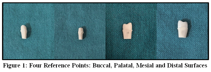 | Figure 1: Four Reference Points: Buccal, Palatal, Mesial and Distal Surfaces
|
For the conventional group, impressions were made using polyvinyl siloxane impression material according to the manufacturer’s instructions and for the digital groups, impressions were obtained using two different intraoral scanners, andDifferent model materials, Type IV die stone (Neelkanth, India) for conventional and 3D-printed resin (ELEGOO, China) fordigital groups were used as per workflow requirements.The 3D printing process was performed using ELEGOO printer with a layer thickness of 50 µm, followed by post-curing as per manufacturer’s instructions to ensure dimensional accuracy. This different model materials may introduce a potential confounding variable, and its possible influence on marginal fit has been considered during interpretation All models were subsequently scanned using a laboratory scanner (Shining 3D, China) after application of scan spray (Alpha Dent, Korea) to enhance scanning accuracy and surface detail capture.
Monolithic zirconia crowns were designed using CAD software (EXOCAD, Germany) with standardized parameters(Figure 2), including an internal thickness of 0.6 mm and a cement space of 0.05 mm. The crowns were milled from pre-sintered zirconia blanks (Nobilcam, China) using a 5-axis milling machine (Roland DWX-53DC, Japan). Following milling, the crowns were sintered in a zirconia sintering furnace (LNY model 5F, China) according to the manufacturer’s instructions. Finishing and polishing procedures were carried out to obtain clinically acceptable crowns with standardized surface characteristics (Figure 3).
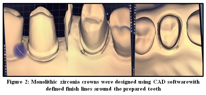 | Figure 2: Monolithic zirconia crowns were designed using CAD softwarewith defined finish lines around the prepared teeth.
|
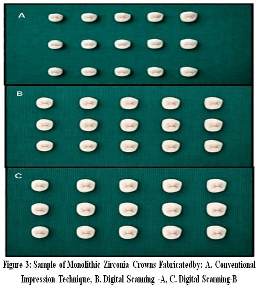 | Figure 3: Sample of Monolithic Zirconia Crowns Fabricatedby: A. Conventional Impression Technique, B. Digital Scanning -A, C. Digital Scanning-B
|
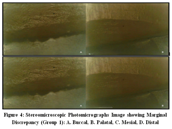
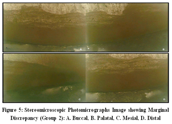
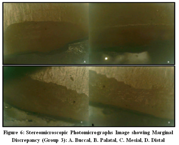
Marginal adaptation of all fabricated crowns was evaluated at the four predetermined reference points using a stereomicroscope at 45× magnification.Marginal fit was evaluated without cementation to eliminate variability introduced by cement thickness. Standardized digital images were captured and analyzed using image analysis software to measure marginal gap values in micrometers (µm). Consistent imaging conditions were maintained throughout the evaluation process to ensure accuracy andreproducibility of measurements (Figure 4,5,6).
| Figure 4: Stereomicroscopic Photomicrographs Image showing Marginal Discrepancy (Group 1): A. Buccal, B. Palatal, C. Mesial, D. Distal
|
| Figure 5: Stereomicroscopic Photomicrographs Image showing Marginal Discrepancy (Group 2): A. Buccal, B. Palatal, C. Mesial, D. Distal
|
| Figure 6: Stereomicroscopic Photomicrographs Image showing Marginal Discrepancy (Group 3): A. Buccal, B. Palatal, C. Mesial, D. Distal
|
Data were tabulated using Microsoft Excel 2010 and analyzed using SPSS version 27.0. Descriptive statistics included mean and standard deviation. Normality was assessed using the Kolmogorov-Smirnov test.For normally distributed data, t-test and one-way ANOVA were applied, followed by Tukey’s post hoc test; for non-normal data, Wilcoxon signed-rank and Kruskal–Wallis tests were used. Statistical significance was set at p < 0.05.
Results
This study evaluated the marginal adaptation of monolithic zirconia crowns fabricated using three different impression techniques: conventional impression technique, digital scanner-A(UPCERA, China), and digital scanner-B (Medit i600, South Korea). Table 1 shows intragroup comparison of Marginal discrepancy within eachtechnique and was measured at the buccal, palatal, mesial, and distal surfaces.For the conventional impression group, the mean marginal discrepancy was 77.33 ± 4.386 µm, 80.80 ± 4.074 µm, 84.00 ± 4.174 µm, and79.53 ± 4.340 µmon buccal, palatal, mesial and distal surface respectively, with the mesial surface showing the highest marginal discrepancy.For the digital scanner-A (UPCERA, China), the mean marginal discrepancy values were markedly lower, with 41.60 ± 2.873 µm,44.60 ± 2.873 µm, 46.60 ± 2.873 µm, and 43.60 ± 2.873 µm at the buccal, palatal, mesial and distal surface respectively.Similarly, for the digital scanner-B (Medit i600, South Korea), the mean marginal discrepancy was 42.60 ± 2.873 µm, 45.60 ± 2.873 µm, 47.60 ± 2.873 µm, and 44.60 ± 2.873 µm atbuccal, palatal, mesial and distal surface respectively.Table 2 shows Intragroup analysis demonstrated that, within each technique, the mesial surface consistently exhibited the highest marginal discrepancy, while the buccal surface showed the lowest marginal discrepancy, indicating surface-wise variation in marginal adaptation.The post hoc comparison of marginal discrepancies at all four surfaces (buccal, palatal, mesial, and distal) demonstrates a consistent pattern. The conventional impression technique showed higher marginal discrepancy across all surface compared to both digital scanning methods Scanner A(UPCERA, China)and Scanner B(Medit i600, South Korea). All these differences were highly significant (p < 0.001). In contrast, the comparison between Scanner A (UPCERA, China) and Scanner B (Medit i600, South Korea) revealed minimal mean differences of approximately ?1.00 µm across all surfaces, which were statistically non-significant (p > 0.05). This indicates that both digital scanning systems performed similarly and provided superior marginal fit compared to the conventional technique across all evaluated surfaces.
Table 1: Intragroup Comparison of Marginal Discrepancy of Monolithic Zirconia Crowns Fabricated by Conventional Impression and Digital Scanning Techniques
Parameters | Groups | Mean (µm) | Std. Deviation (µm) | F-value (µm) | p-value, S/NS |
BUCCAL | Conventional | 77.33 | 4.386 | 521.142 | <0.001, HS |
Scanner A | 41.60 | 2.873 | |||
Scanner B | 42.60 | 2.873 | |||
PALATAL | Conventional | 80.80 | 4.074 | 577.654 | <0.001, HS |
Scanner A | 44.60 | 2.873 | |||
Scanner B | 45.60 | 2.873 | |||
MESIAL | Conventional | 84.00 | 4.174 | 602.053 | <0.001, HS |
Scanner A | 46.60 | 2.873 | |||
Scanner B | 47.60 | 2.873 | |||
DISTAL | Conventional | 79.53 | 4.340 | 533.035 | <0.001, HS |
Scanner A | 43.60 | 2.873 | |||
Scanner B | 44.60 | 2.873 |
Table 2: Intergroup Comparison of Marginal Discrepancy of Monolithic Zirconia Crowns Fabricated by Conventional Impression and Digital Scanning Techniques
Dependent Variable | (I) GROUP (µm) | (J) GROUP (µm) | Mean Difference (I-J) (µm) | p-value, S/NS |
BUCCAL | Conventional | Scanner A | 35.73333* | <0.001, HS |
Scanner B | 34.73333* | <0.001, HS | ||
Scanner A | Scanner B | -1.00000 | 0.709, NS | |
PALATAL | Conventional | Scanner A | 36.20000* | <0.001, HS |
Scanner B | 35.20000* | <0.001, HS | ||
Scanner A | Scanner B | -1.00000 | 0.690, NS | |
MESIAL | Conventional | Scanner A | 37.40000* | <0.001, HS |
Scanner B | 36.40000* | <0.001, HS | ||
Scanner A | Scanner B | -1.00000 | 0.696, NS | |
DISTAL | Conventional | Scanner A | 35.93333* | <0.001, HS |
Scanner B | 34.93333* | <0.001, HS | ||
Scanner A | Scanner B | -1.00000 | 0.706, NS |
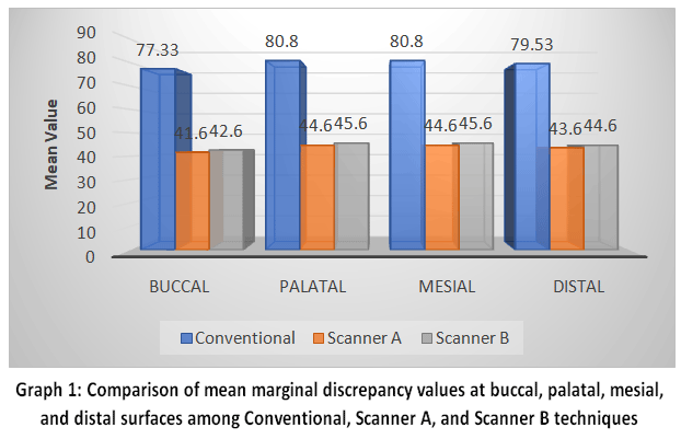
Graph 1 illustrates the comparative mean marginal discrepancy values (µm) of monolithic zirconia crowns fabricated using the conventional impression technique, digital scanner-A (UPCERA, China), and digital scanner-B (Medit i600, South Korea)at the buccal, palatal, mesial, and distal surfaces. The graph clearly demonstrates that the conventional impression technique recorded the highest marginal discrepancy values across all evaluated surfaces, ranging approximately from 77 µm to 84 µm, indicating comparatively poorer marginal adaptation.In contrast, both digital scanning techniques showed substantially lower marginal discrepancy values. Digital scanner-A (UPCERA, China) exhibited values ranging from approximately 41 µm to 47 µm, while digital scanner-B (Medit i600, South Korea) showed values between 42 µm and 48 µm, indicating improved marginal accuracy. Among the two digital systems, scanner-A (UPCERA, China) showed slightly lower marginal discrepancy values compared to scanner-B (Medit i600, South Korea)Post hoc pairwisetest showed that the conventional impression technique had significantly higher marginal discrepancy than both digital scanner-A and scanner-B (p < 0.001). However, no statistically significant difference was observed between the two digital scanners (p > 0.05), indicating comparable performance. Across all three techniques, the mesial surface consistently demonstrated the highest marginal discrepancy, whereas the buccal surface showed the lowest values, indicating better marginal adaptation at the buccal region. Overall, the graph highlights the superior marginal fit obtained with digital impression techniques compared to the conventional method.
| Graph 1: Comparison of mean marginal discrepancy values at buccal, palatal, mesial, and distal surfaces among Conventional, Scanner A, and Scanner B techniques
|
Discussion
This study evaluated the marginal fit of monolithzirconia crowns fabricated using a conventional impression technique was compared with those fabricated using two different digital scanning technique. The results demonstrated clear differences in marginal discrepancy values among the three groups, highlighting the significant influence of the impression technique on marginal accuracy.
Crowns fabricated using the conventional impression technique exhibited the highest marginal discrepancy values at all evaluated reference points. Mean values ranged from approximately 77 µm to 84 µm, with the mesial surface consistently showing the greatest discrepancy. These findings can be attributed to the inherent limitations of conventional impression procedures, including impression material shrinkage, tray distortion, incomplete seating, and dimensional changes during impression removal, stone pouring, and die trimming. Each of these steps introduces potential sources of error, which may accumulate and adversely affect the final marginal fit of the restoration.
In contrast, crowns fabricated using digital scanner-A (UPCERA, China) and digital scanner-B (Medit i600, South Korea) demonstrated significantly lower marginal discrepancy values across all surfaces, with mean values ranging from approximately 41 µm to 47 µm. The superior marginal adaptation observed in the digital groups may be explained by the elimination of multiple intermediate steps associated with conventional impressions.The improved marginal fit observed in digital workflows may be attributed to elimination of intermediate steps, reduced material distortion, and enhanced precision of CAD/CAM systems, leading to improved dimensional stability. Furthermore, the ability to visualize scan data immediately allows for the detection and correction of errors during the scanning process itself, improving overall accuracy.11,12
These findings are consistent with recent studies. Dogan et al., (2025) reported that CAD-CAM fabricated monolithic zirconia crowns demonstrate superior marginal and internal fit due to improved material processing and digital accuracy. Similarly, Seshan et al., (2024) showed that crowns fabricated using digital impressions exhibited better marginal adaptation compared to indirect workflows, emphasizing the role of direct data capture in minimizing distortion. Furthermore, a systematic evaluation by Rojas-Rueda et al., (2025) concluded that digital impressions provide higher accuracy and consistency, supporting their increasing adoption in prosthodontics.13-15
Statistical analysis revealed a highly significant difference between the conventional impression technique and both digital scanning techniques at all reference points, confirming the superior marginal accuracy of digitally fabricated monolithic zirconia crowns. These findings are consistent with previous studies reporting improved marginal adaptation with digital impression workflows.When comparing the two digital scanning systems, digital scanner-A (UPCERA, China) showed slightly lower marginal discrepancy values than digital scanner-B (Medit i600, South Korea)however, this difference wasnot statistically significant.
Surface-wise analysis revealed that the mesial surface exhibited higher marginal discrepancy values across all groups, likely due to its complex anatomy and limited accessibility, which can compromise both impression making and scanning accuracy. Conversely, the buccal surface consistently showed the lowest marginal discrepancy values, possibly due to better visibility and ease of access.This study has limitations, including its in-vitro design, absence of cementation, use of different model materials, and possible operator-related variability.however future scope relies on modifications and standardization for the same.
Conclusion
This study shows that impression technique plays a crucial role in the marginal fit of monolithic zirconia crowns. Conventional methods resulted in greater and uneven discrepancies, while digital scanners produced more accurate and uniform results within clinically acceptable limits. A highly significant difference was found between conventional and digital techniques, with no significant difference between the two digital systems. Overall, digital impressions offer better accuracy and reliability for fixed prosthodontic applications.
Acknowledgement
The authors would like to acknowledge the support of the research team and laboratory staff who contributed to the fabrication of samples and data collection for this study. Their technical assistance and guidance were invaluable in completing this research work.
Funding Sources
The author(s) received no financial support for the research, authorship, and/or publication of this article.
Conflict of Interest
The author(s) declares no conflict of interest.
Data Availability Statement
The manuscript incorporates all datasets produced or examined throughout this research study.
Ethics Statement
Not applicable (This is an in vitro study and does not involve human participants or animals)
Informed Consent Statement
Not applicable
Clinical Trial Registration
Not applicable.
Permission to Reproduce Material from Other Sources
Not applicable.
Author Contributions
Komal Shah: Study design, data collection, analysis, manuscript drafting.
Mukesh Kumar Goyal: Supervision, study design, data interpretation, revision.
Geeta Paul: Data analysis, manuscript revision.
Isha Saxena: Data collection, sample preparation, literature review.
Madhav: Methodology, data validation, editing.
Shreysha: Statistical analysis, proofreading, final preparation.
Nidhi Shree: Data collection, literature review, editing.Dr. Bajaj Amitkumar Shamlal – Supervision, data interpretation, critical review.
References
- Felton D.A., Kanoy B.E., Bayne S.C., Wirthman G.P. Effect of crown margin discrepancies on periodontal health. J Prosthet Dent. 1991;65(3):357–364.
CrossRef - Sorensen J.A. A rationale for comparison of plaque-retaining properties of crown systems. J Prosthet Dent. 1989;62(3):264–269.
CrossRef - Holmes J.R., Bayne S.C., Holland G.A., Sulik W.D. Considerations in measurement of marginal fit. J Prosthet Dent. 1992;67(5):594–599.
CrossRef - Hung S.H., Purk J.H., Tira D.E., Eick J.D. Accuracy of one-step versus two-step putty-wash addition silicone impression techniques. J Prosthet Dent. 1992;68(4):583–589.
CrossRef - Millar B.J., Deb S. Patient experience and acceptability of conventional vs digital impressions. Br Dent J. 2012;213(3):111–114.
- Joda T., Brägger U. Patient-centered outcomes comparing digital and conventional workflows. J Prosthet Dent. 2016;116(4):541–545.
- Sulaiman T.A. Materials in digital dentistry: A review. J Esthet Restor Dent. 2020;32(2):171–181.
CrossRef - Zhang Y., Lawn B.R. Novel zirconia materials in dentistry. J Dent Res. 2018;97(2):140–147.
CrossRef - Nawafleh N., Mack F., Evans J., Mackay J., Lynch E. Accuracy and reliability of methods to measure marginal adaptation. J Prosthodont. 2013;22(4):296–306.
CrossRef - Andriessen F.S., Rijkens D.R., van der Meer W.J., Wismeijer D.W. Applicability and accuracy of an intraoral scanner for digital impressions. Clin Oral Investig. 2014;18(2):521–526.
- Mangano F.G., Hauschild U., Veronesi G., et al. Trueness and precision of four intraoral scanners. BMC Oral Health. 2017;17(1):149.
CrossRef - Michelinakis G., Apostolakis D., Kamposiora P., Papavasiliou G., Özcan M. Digital vs conventional impressions: systematic review and meta-analysis. J Prosthodont. 2021;30(6):560–579.
- Eshan R.R., et al. Comparative evaluation of marginal fit of CAD-CAM crowns fabricated using direct and indirect digital impressions. J Clin Dent. 2024;27(20):1–6.
- Rojas-Rueda S., et al. Accuracy of digital impressions and marginal adaptation of zirconia restorations: a systematic evaluation. J Prosthodont. 2025; 3(2):706–713.
- Dogan D.G., YalugOS. Marginal and internal fit of monolithic CAD-CAM zirconia crowns with varying yttria content and finish line configurations: an in vitro study. Appl Sci. 2025;15(23):12440.
CrossRef


