.jpg)
Immunohistochemical Evaluation of Gingival CD4+ lymphocytes in Response to Allograft Bone Powder (Ceno Bone) - Animal Study
Hadis Moradi  , Ahmad Motaghi* , Atousa Aminzadeh , Alireza Sadighi and Mohammad Hossein Shafazand
1School of Dentistry, Islamic Azad University Isfahan (Khorasgan) Branch, Isfahan, Iran .
http://dx.doi.org/10.12944/EDJ.03.01.05
CD4 T lymphocytes play a central role in allergic reactions. Thus the present study aimed to, immunohistochemically, evaluate the presence of these lymphocytes in rabbit gingival tissues after the replacement of Cenobone. This experimental one way blinded study was performed on 20 gingival tissues gathered from disease-free rabbits with or without bone powder, respectively groups A and B. Immunohistochemical envision method was performed for mapping CD4 lymphocytes. The number and intensity of staining were compared between groups in 5 consequent HPF without overlap with the light microscope in connective tissue. Data were analyzed by Fisher exact test, Wilcoxon, and chi-square statistically in SPSS20 software. The number of CD4 T cells was higher in group A compared to group B.(P=0.02) Pattern of distribution in connective tissue did not show a difference between the two groups. (P=0.41). Results of the present study might confirm the role of CD4 T in an allergic reaction to bone powder material and suggest this cell as a useful factor for the prediction of allergic reactions in the first weeks of surgery. Further studies in this field are required.
Animal; Bone Substitutes; CD4 Positive T-lymphocytes; Graft Rejection; Graft Survival
Copy the following to cite this article:
Moradi H, Motaghi A, Aminzadeh A, Sadighi A, Shafazand M. H. Immunohistochemical Evaluation of Gingival CD4+ lymphocytes in Response to Allograft Bone Powder (Ceno Bone) - Animal Study. Enviro Dental Journal 2021; 3(1). DOI:http://dx.doi.org/10.12944/EDJ.03.01.05
Copy the following to cite this URL:
Moradi H, Motaghi A, Aminzadeh A, Sadighi A, Shafazand M. H. Immunohistochemical Evaluation of Gingival CD4+ lymphocytes in Response to Allograft Bone Powder (Ceno Bone) - Animal Study. Enviro Dental Journal 2021; 3(1). Available From : https://bit.ly/2VsAklV
Download article (pdf) Citation Manager
Introduction
Today dental implants are very popular among populations.1 After tooth extraction and before implant replacement, one main problem is the quantity and quality of remaining bone tissue for future support of the implant 2. So, today bone grafts are used widely in dental science for promoting the healing process of hard tissue and its subsequent healing effects on soft tissue, especially in dental implant surgeries. These grafts can be obtained from patients own body, individuals other than the patient, non-human species, or synthetic man-made products.3,4
Some disadvantages have been reported for autogenous bone grafts like root resorption or feeling discomfort in donor body site. On the other hand, decalcified freeze-dried bone allografts (DFDBA) have shown promise in the bone healing process. So DFDBA are used generally by dentists for bone augmentation. 2 These bone fillers are believed to have no antigenic effect. 3,5 Although in some studies adverse reactions to allogenic bone grafts have been reported. 6,7,8 So a predictive factor for early recognition of allergic reaction and prevention of its progress seems necessary.
The purpose of this study was to try to address a particular factor that might be useable for the prediction of onset or progression of allergic reactions in response to bone powder Ceno-bone (made by HamanandSazBaft Tissue Regeneration Corporation,Iran). 4 Immunological reactions can happen as a humoral response (type I, II, and III reactions) or delayed type IV cell-mediated response. Type IV hypersensitivity is reported to be associated with implant-related allergic reactions. 9,10 In this type of reaction, T cells play a major role. Li et al have reported an increase in CD4+ lymphocyte count in peripheral blood one to two weeks after replacement of decalcified freeze-dried bone scaffolds. 11 Therefore in this study presence of CD4 positive T lymphocytes in rabbit gingival tissues after replacement of Ceno-bone were immunohistochemically investigated as a sign for allergic reaction.
Materials and Methods
This experimental study was performed on 20 gingival tissues obtained from 10 disease-free rabbits. This study was approved by Ethical Committee od Isfahan Azad university. Under general anesthesia, a one-centimeter incision was performed on the right side of mandibular posterior gingival tissue. An equal amount of Ceno bone powder (500 -1000 micromilimetre in total dimension) was placed in the incision area and sutured with vicryl 4-0 (group A). Then on the left side of the mandibular posterior gingival same incision was performed but only sutured with vicryl 4-0 to make sure if the allergic reaction is due to bone powder and not the sutures (group B). (fig1)
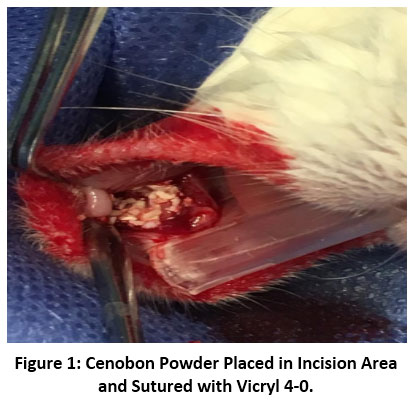 |
Figure 1: Cenobon Powder Placed in Incision Area and Sutured with Vicryl 4-0. Click here to view Figure |
All the rabbits were kept in the same place with the same diet for 20 days. 11 Rabbits with any sign of infection were excluded from the study.
After 20 days a biopsy was performed on the surgical site with a tissue punch. Specimens were placed in 10% buffered formalin and submitted to the pathology laboratory for immunohistochemical staining and mapping of CD4+ lymphocytes. Routine laboratory procedures were performed for each sample and a paraffin blocks were obtained. From each paraffin block, a section (5 microns) was prepared and slides were stained with CD4+ antibody (BiojnexClon, USA) by Envision technique. CD4+ cells were stained brown in light blue background. (fig2 &3)
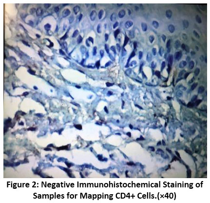 |
Figure 2: Negative Immunohistochemical Staining of Samples for Mapping CD4+ Cells. (×40) Click here to view Figure |
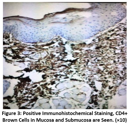 |
Figure 3: Positive Immunohistochemical Staining, CD4+ Brown Cells in Mucosa and Submucosa are Seen. (×10) Click here to view Figure |
The quantity (number of cells) and quality (staining intensity) of stained CD4+ lymphocytes were evaluated using light microscopy in 5 consequent high power fields(HPFs) without overlapping. The pattern of cell distribution was studied in four areas: Junction of epithelium and connective tissue, superficial or papillary connective tissue, deep or reticular connective tissue, and inside the vascular lumen. Data were analyzed by Fisher's exact, Chi-square, and Wilcoxon statistical tests in SPSS20 software.
The Number of Cells
No brown cells -
0-10 cells +
11-30 cells ++
>30 cells +++
Staining Intensity
No brown cells -
Light brown cells +
Dark brown cells ++
Black cells
+++
Results
Wilcoxon test showed a significant difference in the number of CD4+ cells between groups (A: with bone graft and B: without bone graft). (P=0.02) (table1)
Table 1: Number (Percent) of Stained Cells between Two Groups.
|
Number of Stained Cells |
group A n(%) |
group B n(%) |
P-value |
|
0-10 |
2(22.2) |
7(77.8) |
0.02 |
|
11-30 |
1(11.1) |
1(11.1) |
|
|
>30 |
6(66.7) |
1(11.1) |
No difference was observed between the intensity of staining between groups. (Pvalue=0.41) (fig4)
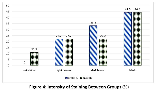 |
Figure 4: Intensity of Staining Between Groups (%) Click here to view Figure |
No difference was observed between the distribution of CD4+ cells in location (superficial or deep connective tissue, junction of epithelium and connective tissue and inside vascular lumen as shown in table 2. (table2)
Table 2: Distribution of Cd4+ Cells between Two Groups (P value > 0.05)
|
Epithelium connective tissue junction |
Cells inside vascular lumen |
Superficial and deep connective tissue |
Deep connective tissue |
|
|
GroupA |
6 |
1 |
9 |
0 |
|
GroupB |
4 |
3 |
8 |
1 |
|
P value |
0.4 |
0.5 |
||
Discussion
T cells are related to rejecting or tolerating the biomaterials and grafts. 12 Anderson et al believe macrophages play an important role in the rejection of biomaterials. 13 Callaghan et al believe this process is set off by CD4 T cells. 10 Wu et al showed an increase in the number of CD4 T-cells 14 days after replacement of bone powder followed by a decrease in the number of these cells in peripheral blood after the second week of surgery. 8 li et al showed CD4 T cells increase in peripheral blood cell count in weeks 1and 2 and a decrease in week 4 after surgery. 11. This increase in the number of CD4 T cells in peripheral blood might be related to systemic or other local factors thus in the present study we tried to evaluate CD4 T cell count directly in the surgical site of bone powder replacement as our study group and the results were compared to a control group: same surgical site in opposite quadrant of the same animal without bone powder but with vicryl sutures. Because hypersensitivity reactions to any kind of sutures have been reported before. 14 We used vicryl sutures to reduce the possibility of such reactions. 15
In the present study, an increase in CD4 Tcell count in connective tissue 20 days after surgery was observed in the study group compared to the control group. We could not compare the results of the present study to studies of Wu et al, and Li et al as the present study is an immune-histochemical study on tissue sections rather than peripheral blood. 8,11 But the recruitment of CD4 T lymphocytes cells into connective tissue might be responsible for the reduction of the number of these cells in peripheral blood. 16 Further studies are required to evaluate the role of these cells in graft survival.
Conclusion
Results of the present study might confirm the role of CD4 T in an allergic reaction to bone powder material and suggest this cell as a useful factor for the prediction of allergic reaction in the first weeks of surgery. Further studies in this field are required.
Acknowledgment
The author would like to thank, school of dentistry, Islamic azad university Isfahan (khorasgan) branch, Isfahan, Iran for their guidance and support to complete this article.
Funding Source
The author(s) received no financial support for the research.
References
- Elani HW, Starr JR, Da Silva JD, Gallucci GO. Trends in Dental Implant Use in the U.S., 1999-2016, and Projections to 2026. J Dent Res. 2018;97(13):1424–1430.
CrossRef - Abolfazli N, Saleh Saber F, Lafzi A, Eskandari A, Mehrasbi S. A Clinical Comparison of Cenobone (A Decalcified Freeze-dried Bone Allograft) with Autogenous Bone Graft in the Treatment of Two- and Three-wall Intrabony Periodontal Defects: A Human Study with Six-month Reentry. Journal of Dental Research, Dental Clinics, Dental Prospects. 2008;2(1):1-8.
- Kumar P, Vinitha B, Fathima G. Bone grafts in dentistry. J Pharm Bioallied Sci. 2013;5(Suppl 1): S125–S127.
CrossRef - Azimi H, Jalayer T, Babaee H. Radiographic Evaluation of the Inhibitory Effect of Ceno-Bone on Alveolar Bone Resorption after Tooth Extraction. J Res Dent Sci. 2012; 9 (3) :156-160
- Peltier LF, Bickel EY, Lillo R, et al. The use of plaster of Paris to fill defects in the bone. Annals of Surgery. 1957; 146:61–69.
CrossRef - Lee GH, Khoury JG, Bell JE, Buckwalter JA. Adverse reactions to OsteoSet bone graft substitute, the incidence in a consecutive series. Iowa Orthop J 2002; 22:35–38.
- Figueiredo A, Silva O, Cabrita S. Inflammatory reaction post-implantation of bone graft materials. ExpPathol Health Sci. 2012;6(1):15-8.
- Wu J, Wang Q, Fu X, Wu X, Gu C, Bi J, Xie F, Kang N, Liu X, Yan L, Cao Y. Influence of immunogenicity of allogeneic bone marrow mesenchymal stem cells on bone tissue engineering. Cell transplantation 2016 ;25(2):229-42.
CrossRef - Chaturvedi T. Allergy related to dental implant and its clinical significance. ClinCosmetInvestig Dent 2013; 5:57–61.
CrossRef - Callaghan CJ, Rouhani FJ, Negus MC, Curry AJ, Bolton EM, Bradley JA, Pettigrew GJ. Abrogation of antibody-mediated allograft rejection by regulatory CD4 T cells with indirect all specificity. The Journal of Immunology. 2007 Feb 15;178(4):2221-8.
CrossRef - Li Y, Yang Z, Qin T. Changes in peripheral blood T lymphocyte subsets of rabbits in the early stage after transplantation of tissue-engineered bone constituted by biologically-derived scaffold. ZhongguoXiufu Chong Jian waiKezaZhi 2007 Feb;21(2):130-4.
- Issa F, Schiopu A, Wood KJ. Role of T cells in graft rejection and transplantation tolerance. Expert Rev ClinImmunol 2010; 6:155.
CrossRef - Anderson JM, Rodriguez A, Chang DT. Foreign body reaction to biomaterials. seminars in immunology 2008; 20:86-100.
CrossRef - Balamurugan R, Mohamed M, Pandey V, Katikaneni H, Kumar KR. Clinical and histological comparison of polyglycolic acid suture with black silk suture after minor oral surgical procedure. The journal of contemporary dental practice 2012; 13: 521-527 DOI: 10.5005/JP-JOURNALS-10024-1179.
CrossRef - AminiSh, Aminzadeh A, Ganji E, Taghva O. Histological Comparison of Gingival Tissue Reaction to Vicryl and Silk Sutures. J Isfahan Dent Sch 2018; 14(4): 381-389. (article in Persian)
- Becker H, Langrock A, Federlin K. Imbalance of CD4+ lymphocyte subsets in patients with mixed connective tissue disease. Clin Exp Immunol 1992;88(1):91–95.
CrossRef


