.jpg)
The Sella Turcica: A Frontier in Dental Diagnosis- A Review
Nidhi Jayaprakash Shetty*  , Amitha H Anand , Shri Kavya Anand , Sandhya Prabha , Sanjana Aniyoor and Harshitha C Suresh
1Department of Paediatric and Preventive Dentistry, Vokkaligara Sangha Dental College and Hospital, Rajiv Gandhi University of Health Sciences, Bangalore, Karnataka India .
http://dx.doi.org/10.12944/EDJ.07.0102.03
The Sella turcica is an important landmark in cephalometric that also serves as a diagnostic indicator in dentistry. Variations in its size and morphology are linked to disturbances in craniofacial development and are often associated with orofacial syndromes such as cleft lip and palate, Williams syndrome, and craniofacial dysostosis. Radiographic evaluation of the Sella region allows early recognition of these conditions. Thus, the Sella turcica represents a valuable frontier in dental diagnosis, bridging radiology with syndromic craniofacial assessment.
Copy the following to cite this article:
Shetty N. J, Anand A. H, Anand S. K, Prabha S, Aniyoor S, Suresh S. H. The Sella Turcica: A Frontier In Dental Diagnosis- A Review. Enviro Dental Journal 2025; 7(1-2).
DOI:http://dx.doi.org/10.12944/EDJ.07.0102.03Copy the following to cite this URL:
Shetty N. J, Anand A. H, Anand S. K, Prabha S, Aniyoor S, Suresh S. H. The Sella Turcica: A Frontier In Dental Diagnosis- A Review. Enviro Dental Journal 2025; 7(1-2).Available here:https://bit.ly/4o4Z6B2
Download article (pdf) Citation Manager

Introduction
The Sella turcica is a saddle-shaped depression in the sphenoid bone that encloses the pituitary gland within the hypophyseal fossa1 [Fig 1]. It serves as a crucial cephalometric landmark in orthodontics and craniofacial growth evaluation, easily identifiable on lateral cephalograms. Its morphology, size, and variations provide information about skeletal development, craniofacial anomalies, and pituitary pathologies.
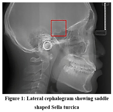 | Figure 1: Lateral cephalogram showing saddle shaped Sella turcica
|
Methodology
A literature search was conducted in PubMed, Scopus, and Google Scholar using keywords such as “Sella turcica morphology,” “dental diagnosis,” “orthodontics,” and “craniofacial anomalies.” English-language human studies relevant to dental or orthodontic diagnosis were included. Titles and abstracts were screened, followed by full-text review, incorporated into the detailed analysis.
Embryological Significance
The Sella turcica region is crucial for the migration of neural crest cells, which move toward the frontonasal and maxillary developmental fields, thereby contributing to midfacial and craniofacial growth.2 The Sella turcica develops in close coordination with the growth of the pituitary gland. Any disturbances in pituitary development may manifest as abnormalities in the Sella turcica morphology.3
Anatomical Features
Sella is located within the body of the sphenoid bone and is bounded anteriorly by the tuberculum sellae, posteriorly by the dorsum sellae, and inferiorly by the pituitary fossa. Its contents include the pituitary gland along with associated vascular and meningeal structures.4
Morphological Variations
The Sella turcica exhibits considerable variation in shape and has been classified according to its radiographic appearance. The commonly recognized forms include circular, oval, flat or shallow, J-shaped, and irregular types, such as notching, bridging, oblique anterior wall, double contour of the floor, and pyramidal dorsum sellae.1
Clinical and Pathological Associations
The size and morphology of the Sella turcica hold significant clinical and pathological relevance, especially concerning the pituitary gland and craniofacial growth. As the pituitary gland resides within the Sella turcica, abnormalities of this gland often present as changes in its dimensions. An enlarged Sella turcica is often associated with conditions like pituitary adenomas, Rathke’s cleft cysts, and intracranial aneurysms, while a reduction in size may occur in hypopituitarism, growth hormone deficiency, or pituitary necrosis. In addition to endocrine disorders, numerous studies have highlighted a strong association between abnormal Sella turcica morphology and a range of craniofacial syndromes and anomalies, such as Williams syndrome, Down syndrome, osteogenesis imperfecta, Cri-du-chat syndrome, trisomy 18, Fragile X syndrome, Axenfeld–Rieger syndrome and cleft lip and palate. These findings highlight the significance of radiographic evaluation of the Sella turcica as a diagnostic marker in paediatric patients with syndromic presentations. In addition, deviations in Sella turcica morphology have been linked with dental anomalies, particularly disturbances in tooth number, shape, and eruption pattern, further emphasizing its clinical importance in orthodontics and paediatric dentistry.5
Radiographic Importance in Orthodontics
Clearly visible on lateral cephalometric radiographs. Serves as a stable reference point (S-point) in cephalometric analyses.
Used in: Sella–Nasion plane (SN plane): essential in evaluating maxillary and mandibular position. Growth prediction studies. Assessment of skeletal and craniofacial anomalies.6
Significance of Sella Turcica
The Sella turcica serves as an important landmark in the fields Paediatric dentistry and orthodontics due to its role as a stable cranial base reference point for cephalometric analysis. Positioned at the centre of the cranial base, it provides a reliable landmark for assessing the anteroposterior and vertical relationships of the jaws. Its evaluation is crucial in determining the growth direction and magnitude of the maxilla and mandible, thereby aiding in treatment planning and growth prediction. Variations in the morphology of the Sella turcica have been shown to correlate with skeletal malocclusions as well as with several craniofacial syndromes, making it an important diagnostic marker.7 In paediatric and syndromic patients, where early detection of anomalies is essential for comprehensive management, the radiographic study of the Sella turcica serves as a valuable diagnostic tool, contributing significantly to both orthodontic evaluation and interdisciplinary care.8
Abnormal Morphologies of Sella Turcica and Their Association with Craniofacial Syndromes
The Sella turcica morphology has been shown to exhibit distinct variations in both normal individuals and patients with craniofacial anomalies. Radiographic assessment of the Sella turcica not only provides information about skeletal and pituitary development but also assists in diagnosing certain craniofacial syndromes.
Several studies, including those by Axelsson et al. (2004), Kucia et al. (2010), and Sathyanarayana et al. (2013), have highlighted a strong association between abnormal Sella turcica shapes and craniofacial syndromes. The following morphological deviations are of clinical significance:
Oblique Anterior Wall – Down Syndrome
Patients with Down syndrome often present with an obliquely angled anterior wall of the Sella turcica [Fig 2]. This morphological change is linked with midfacial hypoplasia and altered cranial base growth patterns commonly observed in these individuals.9
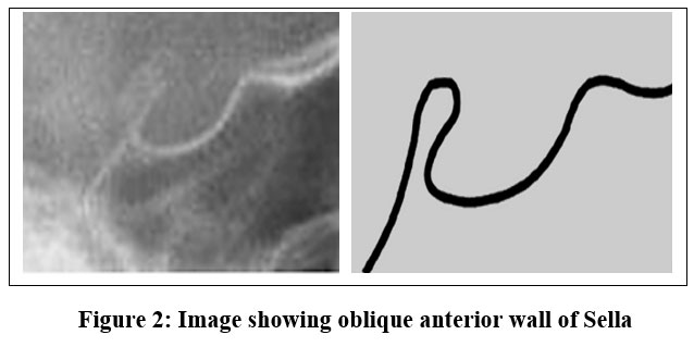 | Figure 2: Image showing oblique anterior wall of Sella
|
Sella Turcica Bridge – Axenfeld-Rieger Syndrome
A Sella turcica bridge [Fig 3], results from ossification connecting the anterior and posterior clinoid processes., is frequently associated with Axenfeld-Rieger syndrome. This syndrome affects ocular, dental, and craniofacial development.10
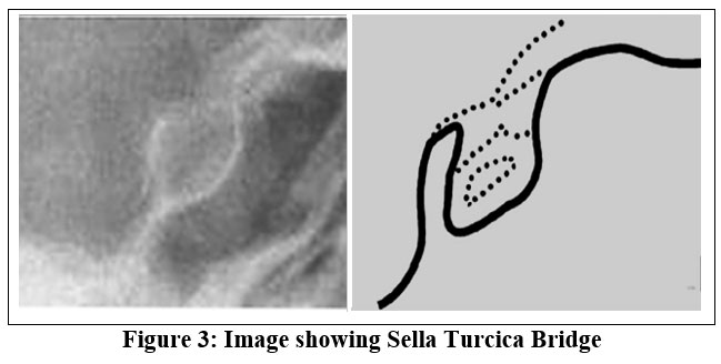 | Figure 3: Image showing Sella Turcica Bridge
|
Irregular Posterior Wall – Apert Syndrome
Apert syndrome, characterized by craniosynostosis and midface hypoplasia, often exhibits an irregular posterior contour of the Sella turcica [Fig 4]. This reflects the disturbed cranial base remodelling process seen in syndromic craniosynostosis.11
 | Figure 4: Image showing Irregular Posterior Wall of Sella
|
Pyramidal Shape – Crouzon Syndrome
A pyramidal configuration of the Sella turcica has been seen in individual with Crouzon syndrome, another craniosynostosis disorder [Fig 5]. This reflects abnormal growth at the Spheno-occipital synchondrosis and distorted cranial base architecture.11
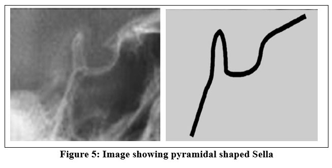 | Figure 5: Image showing pyramidal shaped Sella
|
Hutchinson-Gilford Progeria Syndrome
In Hutchinson-Gilford syndrome/ Progeria, the Sella turcica may exhibit a J-shaped configuration [Fig 6], likely reflect premature skeletal maturation and alter cranial base development.12
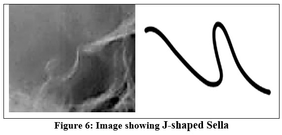 | Figure 6: Image showing J-shaped Sella
|
Uneven Contour of Clivus – Cri-du-Chat Syndrome
Individuals with Cri-du-Chat syndrome have been observed to show an uneven contour of the clivus and Sella floor [Fig 7], consistent with generalized craniofacial dysmorphology.13
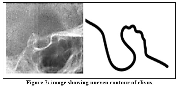 | Figure 7: image showing uneven contour of clivus
|
Clinical Implications
The identification of these abnormal Sella turcica morphologies on routine lateral cephalograms can aid orthodontists in the early recognition of underlying syndromes. Since many of these conditions have dental anomalies and growth disturbances, knowledge of these radiographic markers is vital for treatment planning and interdisciplinary care. Thus, Sella turcica serves not only as a cephalometric landmark but also as a diagnostic indicator of craniofacial syndromes.11
Conclusion
The Sella turcica is not only a key anatomical structure but also a diagnostic window into pituitary health and craniofacial development. Its morphology offers insights into skeletal discrepancies, craniofacial syndromes, and pituitary pathologies. For orthodontists, it remains a fundamental cephalometric landmark for growth assessment, diagnosis, and treatment planning.
Acknowledgement
The authors sincerely thank Dr. Amitha H Anand, Associate Professor, Department of Paediatric and Preventive Dentistry, Vokkaligara Sangha Dental College & Hospital, Bengaluru, Karnataka, India, for her valuable guidance and constructive feedback. We also appreciate Dr. Shri Kavya Anand, Dr. Sandhya Prabha, Dr. Sanjana Aniyoor, and Dr. Harshitha C Suresh, Department of Paediatric and Preventive Dentistry, Vokkaligara Sangha Dental College & Hospital, Bengaluru, Karnataka, India, for their support, encouragement, and constructive feedback throughout the study.
Funding Sources
The author(s) received no financial support for the research, authorship, and/or publication of this article.
Conflict of Interest
The authors do not have any conflict of interest.
Data Availability Statement
This statement does not apply to this article.
Ethics Statement
This research did not involve human participants, animal subjects, or any material that requires ethical approval.
Informed Consent Statement
This study did not involve human participants, and therefore, informed consent was not required.
Clinical Trial Registration
This research does not involve any clinical trials.
Author Contributions
Nidhi Jayaprakash Shetty: Conceptualization, methodology, writing – Original Draft.
Shri Kavya Anand: Analysis, review & Editing.
Sandhya Prabha, Sanjana Aniyoor, Harshitha C Suresh: Review & Editing.
Amitha H Anand: Visualization, supervision and final approval of the manuscript.
References
- Roomaney IA, Chetty M. Sella turcica morphology in patients with genetic syndromes: A systematic review. Orthod Craniofac Res. 2021;24(2):194-205. doi:10.1111/ocr.12426
CrossRef - Mølsted K, Boers M, Kjær I. The morphology of the sella turcica in velocardiofacial syndrome suggests involvement of a neural crest developmental field. Am J Med Genet A. 2010;152A(6):1450-1457. doi:10.1002/ajmg.a.33381
CrossRef - Kjær I. Sella turcica morphology and the pituitary gland—a new contribution to craniofacial diagnostics based on histology and neuroradiology. Eur J Orthod. 2015;37(1):28-36. doi:10.1093/ejo/cjs091
CrossRef - Önal V, Evren A, Chatzioglou GN, Tellio?lu AM. Anatomical features of sella turcica with comprehensive literature review. Rev Assoc Médica Bras. 2023;69(8):e20230402. doi:10.1590/1806-9282.20230402
CrossRef - Gopalakrishnan U, Mahendra L, Rangarajan S, Madasamy R, Ibrahim M. The Enigma behind Pituitary and Sella Turcica. Case Rep Dent. 2015;2015:1-5. doi:10.1155/2015/954347
CrossRef - Sarhan OA. Sella?Nasion linc revisited. J Oral Rehabil. 1995;22(12):905-908. doi:10.1111/j.1365-2842.1995.tb00239.x
CrossRef - Andredaki M, Koumantanou A, Dorotheou D, Halazonetis DJ. A cephalometric morphometric study of the sella turcica. Eur J Orthod. 2007;29(5):449-456. doi:10.1093/ejo/cjm048
CrossRef - Mortezai O, Rahimi H, Tofangchiha M, et al. Relationship of the Morphology and Size of Sella Turcica with Dental Anomalies and Skeletal Malocclusions. Diagnostics. 2023;13(19):3088. doi:10.3390/diagnostics13193088
CrossRef - Korayem M, AlKofide E. Size and shape of the sella turcica in subjects with D own syndrome. Orthod Craniofac Res. 2015;18(1):43-50. doi:10.1111/ocr.12059
CrossRef - Meyer?Marcotty P, Weisschuh N, Dressler P, Hartmann J, Stellzig?Eisenhauer A. Morphology of the sella turcica in Axenfeld–Rieger syndrome with PITX2 mutation. J Oral Pathol Med. 2008;37(8):504-510. doi:10.1111/j.1600-0714.2008.00650.x
CrossRef - Iskra T, Stachera B, Mo?d?e? K, et al. Morphology of the Sella Turcica: A Meta-Analysis Based on the Results of 18,364 Patients. Brain Sci. 2023;13(8):1208. doi:10.3390/brainsci13081208
CrossRef - Ullrich NJ, Silvera VM, Campbell SE, Gordon LB. Craniofacial Abnormalities in Hutchinson-Gilford Progeria Syndrome. Am J Neuroradiol. 2012;33(8):1512-1518. doi:10.3174/ajnr.A3088
CrossRef - Kjær I, Niebuhr E. Studies of the cranial base in 23 patients with cri?du?chat syndrome suggest a cranial developmental field involved in the condition. Am J Med Genet. 1999;82(1):6-14. doi:10.1002/(SICI)1096-8628(19990101)82:1%3C6::AID-AJMG2%3E3.0.CO;2-%23
CrossRef


