Ameloblastomatous Calcifying Odontogenic Cyst: Case Report
Atousa Aminzadeh1 and Alireza Sadighi2
1Department of Oral Pathology, School of Dentistry, Islamic Azad University Isfahan (khorasgan) branch, Isfahan, Iran .
2Department of Oral Surgery, School of Dentistry, Islamic Azad University Isfahan (khorasgan) branch, Isfahan, Iran .
Corresponding author Email: Ar.sadighi@khuisf.ac.ir
DOI: http://dx.doi.org/10.12944/EDJ.01.01.02
Calcifying odontogenic cyst (COC) or calcifying cystic odontogenic tumor was first introduced in 1962 by Gorlin et al., as a possible oral counterpart of calcifying epitheliomas of Malherbe in skin. This lesion is a rare odontogenic lesion with variable clinico-histological characteristics. Three different histologic subtypes has been reported for COC. In this study we presented a female patient diagnosed with ameloblastomatous COC a very rare variant of this lesion and challenges regarding microscopic diagnosis and treatment of it is discussed.
Copy the following to cite this article:
Aminzadeh A. Sadighi A. Ameloblastomatous Calcifying Odontogenic Cyst: Case Report.Enviro Dental Journal 2019; 1(1).
DOI:http://dx.doi.org/10.12944/EDJ.01.01.02Copy the following to cite this URL:
Aminzadeh A. Sadighi A. Ameloblastomatous Calcifying Odontogenic Cyst: Case Report. Enviro Dental Journal 2019; 1(1). Available from: https://bit.ly/2lRJNkU
Download article (pdf) Citation Manager
Select type of program for download
| Endnote EndNote format (Mac & Win) | |
| Reference Manager Ris format (Win only) | |
| Procite Ris format (Win only) | |
| Medlars Format | |
| RefWorks Format RefWorks format (Mac & Win) | |
| BibTex Format BibTex format (Mac & Win) |
Article Publishing History
| Received: | 17-04-2019 |
|---|---|
| Accepted: | 19-06-2019 |
| Reviewed by: | Daisa Pereira |
| Second Review by: | Anka Letic |
| Final Approval by: | Dr. Surinder Pal Singh Sodhi |
Introduction
Calcifying odontogenic cyst (COC) or calcifying cystic odontogenic tumor was first introduced in 1962 by Gorlin et al., as a possible oral counterpart of calcifying epitheliomas of Malherbe in skin.COC is a rare odontogenic lesion with variable clinico-histological characteristics. According to literature only 2% of all odontogenic lesions are COC. Mostly COCs grow as cysts; however in less than 5% COCs can occur as a solid tumor-like mass.1,2
The basic histopathologic criteria introduced for microscopic diagnosis of cystic COC is an odontogenic ameloblastoma-like epithelium containing numerous ghost cells and calcification. (1) But in regard to some microscopic details observed in afew cystic COCs three different histologic subtypes has been considered for cystic COC: simple unicystic type,odontoma producing type and ameloblastomatous proliferating type.(3)65% of cases are simple cystic which is composed of squamous or stellate reticulum like epithelium with or without palisading of basal cells, large ghost cells with eosinophilic cytoplasm which maygo through dystrophic calcification,dentinoid material and melanin deposits. Odontoma associated type actually shows the microscopic features of simple cystic type plus an odontoma associated with it and composes nearly 22% of COCs.Ameloblastomatous type shows stellate reticulum like areas with palisaded basal cells and reverse nuclear polarization and ameloblastoma proliferations in cyst wall.3,4
According to a literiture review in pubmed only afew Ameloblastomatous COCs have been reported which are summerized in table 1.
Table 1: Ameloblstomatous COCs reported in Pubmed
|
Author(s) |
Year |
Country |
Age |
Gender |
Place |
Clinic |
Treatment |
Recurrence |
|
Aithalet al.,5 |
2003 |
India |
28 |
Female |
Mandible |
painless swelling well-defined bony, hard, non-tender swelling of 2.5 cm × 2 cm with smooth surface |
- |
No |
|
Iida et al.,6 |
2004 |
Japan |
17 |
Male |
Mandible |
Painful swelling from second molar to ramus and coronoid process |
Enucleati-on |
No |
|
Kamboj et al.,7 |
2007 |
India |
58 |
Female |
Mandible |
Pain for 5 years and history of swelling for 2 yearsfrom canine to ramus, condyle and coronoid |
Mandibul-ectomy |
No |
|
Singh et al.,8 |
2013 |
India |
24 |
Female |
Mandible |
Swelling for 6 months |
Enucleati-on |
No |
|
Samuel et al.,9 |
2013 |
India |
13 |
Female |
Mandible |
Painless swelling with mild facial asymetry |
Enucleati-on |
No |
|
Menat et al.,10 |
2013 |
India |
20 |
Male |
Mandible |
Swelling for 2 years from mandibular left first molar up to ramus condyle and coronoid |
Hemiman-dibulecto-my with disarticul-ation |
No |
|
Devaraju et al.,2 |
2015 |
India |
65 |
Male |
Mandible |
Painful swelling watery discharge on chewing food |
Not mentioned |
Not evaluated |
|
Bhat et al.,11 |
2015 |
India |
19 |
Male |
mandible |
Swelling extended from premolar to ramus |
Marginal mandibul-ectomy |
NO |
In regard of proper diagnosis and treatment of this lesion the question is how to differentiate it from unicystic ameloblastoma and Ameloblastoma ex coc.And what would be the best treatment planning for this lesion in favor of patient and possibility of recurrence.
In this paper, a case of ameloblastomatous COC is reported which occurred in upper jaw pushing toward maxillary sinus and obliterating its space without bone perforation. In two year follow up after enucleation of cyst no recurrence was observed.
Case Presentation
A 63-year-old female with a two-year history of swelling in her right cheeck and an aspiration biopsy report suggestive of hemangioma was referred to oral maxilofacial surgeon. In extra oral examination, a considerable asymmetry was noticed on the right side of zygomatic area (figure 1).
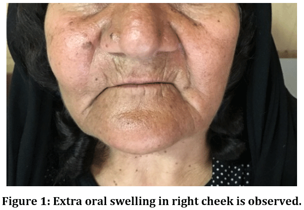 |
Figure 1: Extra oral swelling in right cheek is observed. Click here to view Figure |
Intraoral examination showed an expansion of right maxillary vestibule making her denture hard to fit. The patient had no history of pain or paresthesia. No cervical lymphadenopathy was noticed.
A doppler sonography was performed which denied the vascular nature of lesion.Facial and paranasal CT scan showed fullness and expansion of the lateral and medial wall of right maxillary sinus, but the orbital floor was not elevated. Other sinuses were clear, with no sign of sinus perforation. (Figures 2 ) Her past medical history was irrelevant. No history of allergy or other systemic diseases was reported by the patient.
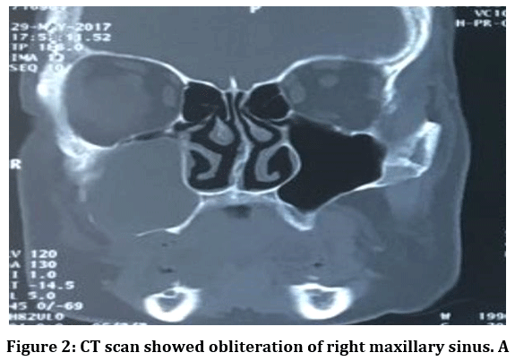 |
Figure 2: CT scan showed obliteration of right maxillary sinus. A Click here to view figure |
With a provisional diagnosis of sinus mucocele and differential diagnosis of odontogenic cysts, a complete excisional surgery under general anesthesia was performed. A brown-gray soft tissue lesion in several pieces some with cystic structure was submitted to pathology laboratory in 10% buffered formalin. In microscopic examinations a cystic lumen lined with columnar basal cells and surface stellate reticulum like cells was observed (Figure3 a,b,c).Numerous ghost cells and spherical calcifications within the ghost cells was seen. A plexiform ameloblastomatous proliferation was observed in the cyst wall with multiple ghost cells. Accordingly a definitive diagnosis of ameloblastomatous COC was made. Patient was under post-operative follow up with no reported complication and recurrence 24 months after surgery.
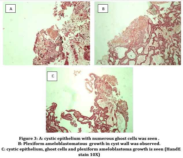 |
Figure 3: A: cystic epithelium with numerous ghost cells was seen. B: Plexiform ameloblastomatous growth in cyst wall was observed. C: cystic epithelium, ghost cells and plexiform ameloblastoma growth is seen (HandE stain 10X) Click here to view Figure |
Discussion
COC represents only one percent of all odontogenic cysts.3 The World Health Organization (WHO)has classified COC as a neoplasm and applied the term calcifying cystic odontogenic tumor for benign cystic types of COC and dentinogenic ghost cell tumor for benign solid-type COCs. Various clinical and histological features have been reported for this lesion. However, no united definition exists regarding the classification of COCs. For many years, the nature of COC resulted in conflicting theories on whether it is a cyst, neoplasm or hamartoma.12
The basic histopathologic criterion introduced for cystic COC is an odontogenic ameloblastoma-like epithelium containing ghost cells which may go through calcification.1
Three different histologic subtypes have been reported for cystic COCs, including simple unicystic type, odontoma producing type and ameloblastomatous proliferating type.3 It is important to differentiate ameloblastomatous COC from unicystic ameloblastoma and ameloblastoma ex COC to prevent any unnecessary invasive treatment procedure. According to a literature review in PubMed, only a few ameloblastomatous COCs have been reported previously, which are summarized in table 1. With regard to the proper diagnosis and treatment of this lesion, the questions that arise are how is it differentiated from unicystic ameloblastoma and ameloblastoma ex COC? and what would be the best treatment plan for this lesion in favor of both patient and possibility of recurrence? In brief would this type of COC have different biological behavior,treatment and prognosis than other histologic subtypes?.3,13,14,15
COCs show nuclear and cytoplasmic positivity for beta catenin.4 But the immune-histochemistry staining is not available all time as it was not available for our patient so a histologic criteria is necessary for differentiating between these similar lesions with different treatment and prognosis. Ameloblastomatous COC shows intra luminal and intramural plexiform (sometimes follicular) ameloblastomatous proliferation admixed with ghost cells and calcification. Unicystic ameloblastoma has both intra-luminal and mural ameloblastomatous proliferation but lacks the ghost cells and calcifications seen in ameloblastomatous COC. Ameloblastoma ex COC shows ameloblastic proliferation within the cyst wall without any ghost cell or calcification.13
Adenoid ameloblastoma and odontogenic carcinoma can as well be considered in differential diagnosis of ameloblastomatous COC. Adenoid ameloblastoma also contains ghost cells and dentinoid similar to ameloblastomatous COC but has a pseudocribriform morphology. Odontogenic carcinoma has the dentinoid material but very few ghost cells and shows infiltration to adjacent stroma.4
In the present case selecting the proper surgical treatment was challenging. In the previously reported cases shown in table 1, ameloblastomatous COCs have been mostly treated by enucleation, hemi- mandibulectomy was reserved for large cases accompanying bone perforation. In our case, although lesion pushed itself toward maxillary sinus, the sinus wall was clear and intact. (figure 4) Thus, it was decided to omit the lesion by enucleation and curettage, with preservation of the infraorbital nerve.
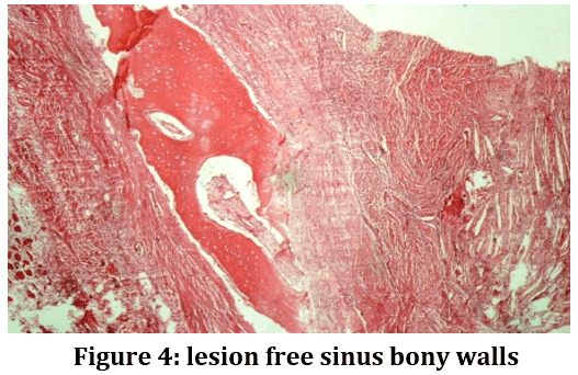 |
Figure 4: lesion free sinus bony walls Click here to view Figure |
In a 24 month follow up, no sign of recurrence was noticed clinically and in CT scan(image not available) (Figure 5). In all the cases reported before, no recurrence was seen, which shows this lesion does not have a neoplastic nature despite the tumoral growth in its wall. The clinical significance of mural ameloblastomatous proliferation in COC cyst wall is still unclear.14 So until today enucleation and close follow remains the best treatment plan for small ameloblastomatous COCs without bone infiltration.
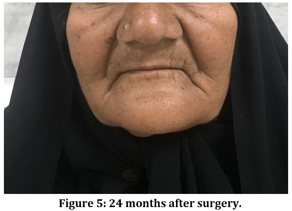 |
Figure 5: 24 months after surgery. Click here to view Figure |
Conclusion
Ameloblastomatous COC should be differentiated from ameloblastoma ex COC, unicystic ameloblastoma and calcifying ghost cell odontogenic tumor to prevent unnecessary invasive approaches. Complete microscopic examination of the entire sample and a careful post operative follow-up is highly recommended.
Acknowledgments
This article was not supported by any and has no conflict of interest.
References
- Neville BW, Damm DD, Chi AC, Allen CM. Oral and maxillofacial pathology: Elsevier Health Sciences; 2015.
- Devaraju RR, Duggi LS, Gantala R, Sanjeevareddygari S, Potturi A. Ameloblastomatous Calcifying Cystic Odontogenic Tumour: A Rare Variant. Journal of Clinical and Diagnostic Research: JCDR. 2015;9(3):ZD20-ZD21. doi:10.7860/JCDR/2015/12600.5717.
CrossRef - Sonone A, Sabane VS, Desai R. Calcifying Ghost Cell Odontogenic Cyst: Report of a Case and Review of Literature. Case Reports in Dentistry. 2011;2011:32874 doi:10.1155/2011/328743.
CrossRef - Woo SB.Oral pathology: A comprehensive atlas and text. 2nd ed. Elsevier hiladelphia. 2017; 411-16.
- Aithal D, Reddy B, Mahajan S, Boaz K, Kamboj M. Ameloblastomatous calcifying odontogenic cyst: a rare histologic variant. Journal of oral pathology & medicine. 2003;32(6):376-8.
CrossRef - Iida S, Ueda T, Aikawa T, Kishino M, Okura M, Kogo M. Ameloblastomatous calcifying odontogenic cyst in the mandible. Dentomaxillofac Radiol 2004 Nov;33(6):409-12.
CrossRef - Kamboj M1, Juneja M. Ameloblastomatous Gorlin's cyst. J Oral Sci 2007 Dec;49(4):319-23.
CrossRef - Singh HP, Yadav M, Nayar A, Verma C, Aggarwal P, Bains SK. Ameloblastomatous calcifying ghost cell odontogenic cyst-a rare variant of a rare entity. Annali di stomatologia 2013;4(1):156.
- Samuel S, Sreelatha S, Venkatesh S, Nair PP. Ameloblastomatous calcifying odontogenic cyst: a rare histological variant. BMJ case reports 2013;2013:bcr2013009137.
CrossRef - Menat S, Md S, Attur K, Goyal K.Case Rep Dent. Ameloblastomatous CCOT: A Case Report of a Rare Variant of CCOT with a Review of the Literature on Its Diverse Histopathologic Presentation 2013;2013:407656.doi: 1155/2013/407656.Epub 2013 Oct 9.
CrossRef - Bhat S, Shetty SR, Babu SG, Shetty P, Ka F.A rare variant of calcifying odontogenic cyst with ameloblastoma presentation.Stomatologija. 2015;17(4):131-4.
- Irani S, Foroughi F. Histologic Variants of Calcifying Odontogenic Cyst: A Study of 52 Cases. The journal of contemporary dental practice. 2017;18(8):688.
CrossRef - Kallalli BN, Rawson K, Patel V, Patil S. Ameloblastomatous calcifying odontogenic cyst: A rare histologic variant. Journal of Indian Academy of Oral Medicine and Radiology 2015;27(1):123.
CrossRef - Yuwanati MB, Tupkari JV, Mhaske S, Avadhani A, Joshi P. Ameloblastomatous calcifying ghost cell odontogenic tumor: A case report. International Journal of Case Reports and Images 2012;3(9):21–25.
- Aparna M, Gupta M, Sujir N, Kamath A, Solomon M, Pai K, et al.,. Calcifying Odontogenic Cyst: A Rare Report of A Nonneoplastic Variant associated with Cholesterol Granuloma. The journal of contemporary dental practice 2013;14(6):1178
CrossRef
.jpg)


